


Microscope Glossary of Terms
May 19, 2021
A
-
Arm – the part of the microscope that connects the eyepiece tube to the base.
- Articulated arm – a type of microscope stand that clamps to or sits on a surface (like a table) and moves in three dimensions.
- Fixed arm – a type of microscope stand with a solid connection to the base. Typically used with low power stereo microscopes.
B
- Balance plate – a plate found on spotting scopes to properly balance the
- Base – the bottom support of the microscope.
- Body – the main section of the microscope, usually including the eyepiece and objective lenses but not the focusing block.
- Brightfield illumination – an illumination technique that yields dark objects on a bright background. The light source is positioned below the sample. It is the opposite of Darkfield Illumination. For more information, click here.
C
- Calibration – a mathematical process of determining the true distance when using an eyepiece reticle.
-
Coaxial focus – a focusing system that has both the coarse and fine focusing knobs mounted on the same axis, allowing differential measurements to be recorded.
- Coarse focus – the focus control knob on the microscope that quickly moves the objective lenses toward or away from the specimen. Works with fine focus.
- Fine focus – the focus control knob on a microscope that fine-tunes the focus on the specimen. It is also commonly used to change the plane of focus for examination of different parts of the specimen. Works with coarse focus.
-
Condenser Lens – a lens mounted in or below the stage (substage) to focus or condense light onto the specimen.
- Abbe condenser – a specially designed substage lens that has an iris type aperture to control the amount of the light that enters the lens system with magnifications above 400x.
-
Contrast – the difference in light intensity between the image and the adjacent background relative to the overall background intensity.
- Contrast plate – a circular, opaque plate that is placed on the stage of a low power microscope. One side is white, the other is black. You can choose which side to use depending on the coloration of your specimen. Contrast plates are found only on stereo microscopes.
- Oblique contrast – a contrast-enhancing optical technique used to increase the contrast of images of thin, transparent, unstained samples, the details of which can be difficult to visualize using brightfield microscopy.
- Phase contrast – a contrast-enhancing optical technique used to enhance the contrast of light microscopy images of transparent and colorless specimens. It enables visualization of cells and cell components that would be difficult to see using an ordinary light microscope.
- Cover slip – a very thin square piece of glass or plastic that is placed over a specimen mounted on a microscope slide to protect the specimen and keep it in place for observation.
D
- Darkfield illumination – an illumination technique that eliminates scattered light from the sample image. The light source is positioned below the sample, but is centrally blocked so that the sample is only illuminated from the sides, thus presenting the sample against a dark background. It is the opposite of Brightfield Illumination. For more information, click here.
- Diaphragm – a disc placed under the stage (substage) to vary the amount of light passing through the stage opening. Helps to properly illuminate the specimen, increase contrast and improve resolution.
- Dimmer – a switch or wheel that adjusts the amount of light coming from the illuminator or light source at the base.
- Diopter adjustment – in binocular microscopes, adjustment of the ring located below or on the eyepiece that allows you to compensate for the difference in eyesight between your right eye and left eye for the sharpest possible image.
E
- Eyepiece lens – the lens that you look into at the top of the microscope. Standard eyepiece lenses usually provide 10x magnification, but some eyepieces provide 5x, 15x, or 20x magnification.
- Eyepiece tube – the tube that houses the eyepiece lens.
F
-
Filters – a circular disk of film or colored glass that attaches to a microscope to enhance details or improve contrast.
- Filter wheel – an accessory to position a selected filter in the imaging path quickly and accurately.
- Field of view (FOV) – in optical microscopes, the area in front of the objective lens assembly that is visible to the user through the eyepiece(s). Additional attachments to the eyepiece(s), such as microscope cameras, may have field of view values of their own that supersede the microscope’s field of view.
- Flange – a projecting flat rim, collar, or rib on an object, serving to strengthen or attach or (on a wheel) to maintain position on a rail.
- Focusing – the process of moving the lens configuration (eyepiece and objective or digital sensor and objective) in relation to the specimen to achieve a sharp image. This could be movement of the stage holding the specimen or movement of the head of the microscope.
H
-
Head – The upper part of the microscope that contains the eyepiece tube and prisms.
- Monocular head – a microscope head with a single eyepiece lens.
- Binocular head – a microscope head with two eyepiece lenses, one for each eye.
- Dual head – a microscope with a single eyepiece lens coming out one side and an additional single eyepiece tube coming either off the top or from the opposite side. Dual head microscopes are great when you want to attach a single eyepiece and an imager to the microscope at the same time.
- Trinocular head – a microscope with two eyepiece lenses (one for each eye) and a third port at the top for a camera.
I
-
Illuminator – the light source for your microscope. Four types are commonly used:
- Tungsten illuminator – a bright and hot light. Not recommended for live specimens.
- Fluorescent illuminator – a cool, bright light.
- LED illuminator – a cool light with a long bulb life.
- Halogen illuminator – a very bright and hot light. Not recommended for live specimens.
- Mirror illuminator – a mirror under the stage that reflects a separate light source up through the opening
- Immersion oil – a special oil used with immersion oil objectives that acts as an air-free bridge between the glass slide and the glass in the lens. Immersion oils come in two types: A and B.
-
Immersion oil Type A: oil thatreduces any tendancy to trap airImmersion oil Type B: oil that is thick enough for viewing multiple slides with one application.
- Inclination joint – the location at which the microscope arm connects to the microscope base. If there is a pin, you can tilt your microscope back at the inclination joint for more comfortable viewing.
- Interpupillary distance (IPD) – in binocular microscopes and stereoscopes, the measurement, or the potential range of adjustment, available between the exit pupils, as measured in millimeters.
L
- Light source – see illuminator.
M
- Magnification – the amount or degree of visual enlargement of an observed object expressed in “X,” which denotes a 100% increase in size per X. : 4X= 400% increase in size.
- Micrometer (micrometre, micron), abbreviated aμm – a metric linear measurement used in microscopy to identify the size of a viewed subject. One micrometer is equal to 0.001 mm, or about 0.000039 inch.
-
Microscope – an optical instrument used for viewing objects, typically under levels of magnification several hundred times the objects’ actual size. There are three basic parts: head, body, arm.
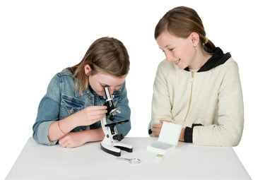
- Compound microscope – a microscope that combines the power of lenses and light to enlarge the subject being viewed. Compound microscopes are among the most common microscopes, often found in science and biology classrooms.
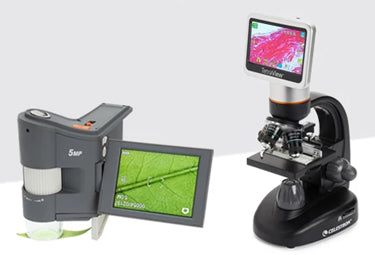
- Digital hybrid microscopes – a microscope that combines parts of a traditional compound microscope with digital components, especially digital viewing and recording heads that either replace or augment traditional eyepieces.
- Electron microscope – a powerful, laboratory grade microscope that allows researchers to view specimens at nanometer size.
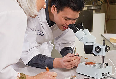
- Stereo microscope – a microscope, generally of less than 80x magnification, that combines two optical paths at slightly different angles, enabling the image to be viewed three-dimensionally under the lenses. Also called a dissecting microscope.
- Pocket microscope – a portable handheld microscope to use in the field.
- Portable microscope – a microscope intentionally designed to be easily moved about and used effectively in a variety of locations beyond a classroom or lab.
-
Mirror – a tool that directs ambient light up through the hole in the stage and illuminates the specimen.
- Flat mirror – a mirror that reflects light evenly upon a specimen to provide a balanced level of illumination.
- Concave mirror – a mirror that concentrates the light and provides brighter illumination at a focused location.
N
- Nosepiece – the part of the microscope that holds the objective lenses, also called a revolving or objective nosepiece or turret.
- Numerical aperture (N.A.) – a number that expresses the ability of a lens to resolve fine detail in an object being observed.
O
-
Objectives standard – the official standard to which a given objective lens or lens assembly was created.
- DIN objectives – abbreviation of “Deutsch Institute fur Normung,” refers to a microscope with a 160mm tube length.
- JIS objectives – abbreviation of “Japanese Industrial Standard,” refers to a microscope with a 170mm tube length.
- RMS objectives – abbreviation of “Royal Microscopical Society,” refers to the standard mounting thread of 0.7965" x 36TPI
-
Objective lens – the lens closest to the object being examined. Objectives are fitted into the nosepiece, and most microscopes have more than one.
- Achromatic objective lens – an objective lens that is corrected for axial chromatic aberration in two wavelengths (blue and red; approximately 486 and 656 nanometers, respectively) that are brought into a single common focal point. Achromatic objectives are also corrected for spherical aberration in the color green (approximately 546 nanometers).
- Apochromatic objective lens – the most highly corrected microscope lens commonly available. Apochromats are chromatically corrected for three colors (red, green, and blue), which largely eliminates all chromatic aberration. They are also spherically corrected for either two or three different wavelengths.
- Semi-plan (semi-planar) objective lens – a lens that provides an approximately 80% flat, clearly focused area in the center of the field of view.
- Oil immersion lens – an objective lens designed to work with a drop of special immersion oil placed between it and the slide.
- Objective nosepiece – see nosepiece.
- Ocular – see eyepiece.
P
- Parcentered – an object that stays in the center of view when the objective is rotated.
- Parfocal – a microscope that, once focused with one objective, remains in focus when the objective is rotated.
- Pointer – a piece of wire that sits in the eyepiece and enables a viewer to point at a specific area of a specimen. By turning the eyepiece, you can adjust the pointer’s position within the field.
R
- Rack and pinion – a type of linear gear system on a microscope that moves an object to your desired viewing position.
- Rack stop – a small screw that does not allow the stage to move too close to the objective lenses, keeping the microscope slide at a safe distance from the objectives.
- Resolution – the level of clarity/detail present in an image. In digital microscopy, the resolution refers to the amount of pixels on the sensor, and is expressed in MP (megapixels).
- Reflection light – existing light or light from an illuminator above the specimen that is reflected up to the microscope’s objective.
- Reticle – a small etched circular glass that is inserted in a microscope eyepiece lens. Reticle patterns include rulers, grids, crossed scales, concentric circles, comparators, grain sizing, and many more.
- Revolving nosepiece – see nosepiece.
S
-
Slide – a flat or concave glass or plastic rectangular plate that the specimen is placed on.
- Prepared Slides – slides that have been prepared with a specimen, sealed and labeled for future use.
- Slip clutch – a mechanical device that protects the gears of the microscope from being turned past their minimum or maximum.
-
Specimen – the object being examined.
- Live specimen – a sample of a live substance or material for examination or study—typically, animal, plant, etc.
-
Stage – the flat plate where slides are placed for observation.
- Mechanical stage – a mechanism that is mounted on the stage to move the slide around, consisting of a slide holder and two knobs.
- Stage clips – a mechanism on either side of the stage that holds the microscope slide in place.
- Stage drift – the stage’s movement during use. Excessive stage drift makes it difficult to keep the specimen in focus.
- Stage plate – a frosted circular glass plate that fits over the lower illuminator. See also contrast plate.
- Sub-stage – the area below the stage that houses the mirrors, diaphragms, or condensers.
-
Stand – a type of connection between the microscope body and base. There are three main types of microscope stands: the pole/post, the fixed arm, and the universal boom stand.
- Pole/post stand – a type of stand that consists of a single post erecting from the base. The microscope can rotate around the post or move up and down on it.
- Fixed arm stand – see arm (fixed arm).
- Universal boom stand – a type of stand that allows you to move the microscope over a large working area (X, Y and Z axis).
- Subject – see specimen.
T
- T-mount – a system that uses T-threads of metric dimensions to mate cameras to lenses or telescopes.
- Tension adjustment – an adjustment of the focusing mechanism to tighten or loosen the stage to reduce stage drift.
- Transmitted light – light that shines through the specimen.
- Turret – See nosepiece.